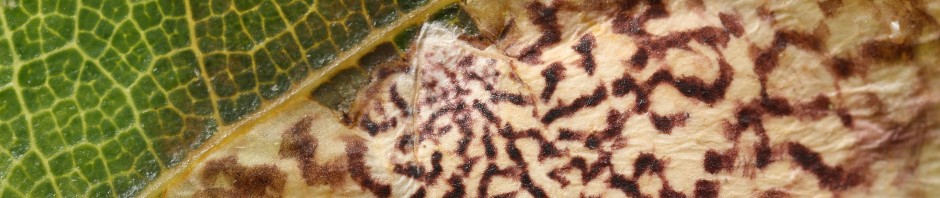
I deliberately waited until I had finished my first book before buying the MP-E 65mm macro lens, because I wanted to focus on making a guide to things that people can actually see. Now that I have it, though, it has enabled me to take animal tracking to a very tiny scale, as illustrated by the photos of agromyzid fly vs. parasitoid wasp exit holes in my previous post. Those are details that my naked eye can still make out, but here is an example of something I never would have noticed without that lens. The caterpillar in the photo below is 5 mm long, the larva of Bucculatrix ivella (Bucculatricidae), one of the few leafminers with a common name: the Groundsel Leaf-perforator Moth. It feeds on groundsel (Baccharis halimifolia), and its name comes from what it does after it is done leafmining. Like most Bucculatrix species (the “ribbed cocoon maker moths”), after making a short leaf mine it emerges and eats little patches out of the leaf surface, usually on the underside. The reason anyone bothered to give it a common name is that it has been introduced as a biological control agent in Australia, where groundsel has become invasive.
Anyway, after I took a few photos of this larva, I zoomed in on the LCD screen to make sure it was in focus, and I noticed a little dot.

See it? Here’s a closer crop of the same photo:

Smack in the middle of the first abdominal segment (the first segment without a leg attached to it) is the hole where a parasitoid wasp inserted her ovipositor. As the larva nonchalantly munched away, a wasp larva was living inside it, biding its time. Not long after I took that photo, the caterpillar spun its cocoon on the side of the vial–you can see why they’re called “ribbed cocoon maker moths”:

It even went to the trouble to build a protective silk fence around itself before spinning its cocoon, to keep away predators (hard to see with the white background; here is a clearer example)–but of course it was too late for that. Once the cocoon was spun, the wasp larva rapidly grew and devoured its host, and eleven days after I collected the caterpillar, this little red braconid emerged–a Polystenidea species, according to Michael Sharkey.


thank for this high magnification macro image of the ‘infected’ larvae. Can you image caapture easily at this high magnification macro…in the field/ in situ? Is this all harvested plant materials that you then setup at your indoor workbench?
These cycles within cycles which you share are profoundly enriching for my outdoors appreciation, Thank you, Charley. charlie guevara/fingerlakes,US
That particular photo was taken indoors, but only because I hadn’t had that camera with me when I collected the larva. There isn’t much difference in the ease of taking these photos indoors vs. outdoors; I don’t use any kind of fancy lighting setup, and my “workbench” consists only of a white dinner plate, which I put inside a large plastic bag (along with the “business end” of the camera) when photographing something that might fly away. The caterpillar photo was taken at 1x, the lowest magnification, and I find it fairly easy to get decent shots that way. At 3-5x it becomes challenging just to find the insect, and when it is in view the depth of field is frustratingly small, but I do what I can.
Charley – I got two very similar Braconids out of the Bucculatrix we collected north of Davis. Nothing else yet, though most had microscopic exit holes already, but I have another half dozen or so that ought to emerge soon (as moths, I imagine).
Cool… I’ve reared many braconids from various microleps and other things, and I don’t remember ever seeing a red one with yellow stigmas like this before.
Pingback: Previews of Parasitoids | BugTracks