Thanks to the fact that I have a searchable email account, and never delete any emails, I was able to quickly reconstruct this timeline just now: On August 1, 2007, I got a reply from Mark Allison, editor at Stackpole Books, to whom I had recently written with a proposal for a field guide to invertebrate tracks and sign, along the same lines as the Bird Tracks & Sign and Mammal Tracks & Sign books that Stackpole has already published, both written by Mark Elbroch. After some discussion and deliberation, on September 10 Mark Allison wrote to let me know Stackpole would like to proceed with the book. As I ramped up my efforts to photograph all manner of curious patterns and objects produced by insects and other invertebrates, I noticed these spots on a hickory leaf on September 30:

Here is a close-up of the pair near the tip of the terminal leaflet, as viewed from the lower surface:

I had no idea what I was looking at. That winter, I spent many days sitting by the shelf where the University of Vermont’s library kept all the insect and spider books, paging through them and making notes about interesting phenomena to look for, as well as getting answers to some of the mysteries I had encountered. Some time over the course of that winter, I read a description that reminded me of these hickory circles. On March 9, 2008, I wrote to Dave Wagner for the first time. After telling him what I was up to, I wrote:
I’m familiar with your excellent caterpillar guides, and I suspect you have answers to a lot of natural history mysteries I’m working on figuring out. But in particular, I’m told you are an expert on leaf miners, so I wanted to see if you can explain the attached image [the same one shown above]. It depicts two blotch mines on a hickory leaf (near Amherst, MA), each with a perfect circle of excrement inside. I have read that the larvae of certain eulophid leaf miner parasites “construct a circle of little faecal pillars about themselves … the larva pupates within this circle and the pillars harden to serve as “pit props”, preventing the collapse of the host mine as the plant tissue dries out.” I’ve never seen this illustrated, but it is tempting to conclude that this is what I’ve photographed. So I’d be grateful for your thoughts on this image, eulophids, and anything else that might make perfect circles in leaf mines.
I’m not sure what the source of that quote was, but I suspect it was something written by S.W. Frost. Now that I have been studying leafminers for many years, I am well acquainted with these fecal pillars, and know that they look nothing like those hickory circles; here is an example of some surrounding the pupal exuviae of Chrysocharis occidentalis in a backlit leaf mine of the beetle Baliosus nervosus (Chrysomelidae):

Sometimes they form more of an oval, but never a perfect circle. Two parallel lines is typical. Anyway, Dave wrote back an hour later:
These don’t look like mines to me. Mines always have parenchyma removed. If you hold the leaf up to the light there should be parenchyma missing. I could be wrong and it is impossible to say from a photograph if backlighting was not used.
My guess is that this is fungal damage and the blemish is more akin to a fairy ring (of mushrooms) than it is to any insect damage, but one a Lilliputian’s scale.
Good luck with your guide.
And I responded,
Thanks for the quick response. I agree that they don’t seem to be mines in the photograph, although I was pretty convinced at the time I took it. If I ever see this sort of thing again, I’ll be sure to reevaluate it, with microscope if necessary. A fungus would certainly explain the lack of larva, pupa, or exit hole.
One of my favorite discoveries at the UVM library was Ray Gagné’s 1989 book, The Plant-feeding Gall Midges of North America. Had I closely studied pages 211-212, I would have found the solution to this riddle there. But it remained a mystery to me when my book was published in April 2010. I’m not sure when I learned that Ray was still active at the Smithsonian, but it was on December 6, 2010 that I first wrote to him with some miscellaneous questions about midge galls. We discussed various things over the next several weeks, and on January 12, 2011, he wrote, “Incidentally, have I sent you a copy of my hickory gall midges paper?” I had not been aware of this paper, and when it arrived in my mailbox 12 days later, I found it was more like a small book (147 pages) than a mere “paper”; it is now available online at the Biodiversity Heritage Library. It features the complex galls of Caryomyia species (Cecidomyiidae), which I wrote about here, and when I perused it I again missed the answer to the hickory circle riddle; it had never occurred to me that these things could be insect galls. In 2011, Ray was working on finishing up a similar publication about the gall midges on hackberries (mostly made by species in the genus Celticecis), and on June 29, I wrote to him:
I’m in Burlington, VT for the week, and I’ve been taking every opportunity to examine hackberry leaves. I finally found some galls yesterday, which I believe are C. spiniformis, and I will send them to you before I head home. Some of them are still very small, so perhaps I will find more later in the season, but clearly diversity and abundance up here are not what they are farther south.
On Monday I found a curious thing on Carya cordiformis [bitternut hickory] leaves, and I wonder if you can offer any insight. The first attached photo shows the appearance of the upper surface of the leaf, a discolored area about 6 mm across. The second shows a dipteran larva feeding on the underside, and the feeding trail glistening with the larva’s slime. I collected leaves with three larvae, and by the end of the day two larvae had gone wandering. The final photo is a close-up of one of these 2-mm larvae. I gave them some moist soil to burrow into, but they both climbed upward in the container, so I’m not sure where they’re trying to go. I would appreciate any thoughts you might have about the identity of these larvae.


Ray’s reply:
The season seems to be late up there for both items that you report on. That you found only spiniformis in Vermont does not surprise me because my co-author on this spent several years at Cornell and never found but that one kind. The other item, on Carya, is Gliaspilota glutinosa that is treated in my hickory paper. These drop to the ground when full grown and remain there until the following year. I don’t know why yours appeared phototropic.
This species was described as Cecidomyia glutinosa by Baron Carl Robert Osten Sacken in 1862. The entire description, which starts at the bottom of p. 193 in this publication, reads:
The small yellowish-orange larva forms no gall, but lives in the open air on the under-side of the leaf, to which it is attached by a viscous substance probably secreted by the leaf. The presence of the larva is indicated on the other side of the leaf by a round yellow spot. The structure of the larva is peculiar: it has rows of fleshy, pointed tubercles along its back, like the larva of C. pini inopis (described below), with which it agrees in some respects in its habit of fastening itself to the surface of the leaf by means of a viscous substance.
I constantly find myself reflecting how much more progress I could be making describing new species if it were still the 19th century, when this was considered an adequate description. Anyway, adults of this species were unknown until April 1995, when Ray Gagné had three females emerge from larvae he had collected in Maryland the previous May. As far as I know these are still the only known adult specimens. It was in the 2008 publication that Ray described the genus Gliaspilota to contain this species. On August 2, 2011, I wrote to him:
I took the attached photos today in Pelham, Massachusetts. I’m sending them as apparent evidence of a second generation of Gliaspilota glutinosa, since your hickory paper states that larvae in the northeastern US have all dropped to the soil by mid-June. I have seen old galls like these many times over the past several years, always appearing as perfect, raised rings, and never would have guessed that they were caused by the same organism that made the meandering trail I asked you about back in June. I wonder what accounts for this variation.


To which he replied:
Fascinating, Charley. I wonder if some of this species just appears later in New England than elsewhere and if the tighter, less viscous ring may be due to leaf senescence. I never found fresh galls this late down here. One way to prove two generations is to gather up a lot of larvae early in the year and see if adults come out in the summer. Otherwise, we’d just be speculating. We’ll have to keep an open mind on this.
I still have not succeeded in rearing adults of this species, but I have only tried a few times. I’ve given you all of this background just to introduce you to the creature that is the subject of this little series I found as I was sorting through my photos from 2025 over the past couple of weeks. On August 7 I was walking in the woods with bryologist Bill Buck and botanist/coleopterist Rob Naczi; Rob and I were helping Bill with his ongoing biodiversity inventory of the town of Kent, New York. I spotted some evidence of Gliaspilota glutinosa, another new species for the town list, and I pointed out one “gall” that still had a larva on it. I was already moving on to look for more species to add, but Bill picked the leaf to take a look at the larva with his hand lens. He asked about what seemed to be another insect sitting on top of the larva. Rob and I each squinted at it through our own hand lenses, and there did seem to be a tiny tiny yellow wasp there, which I imagined was a parasitoid in the process of ovipositing. I only had my phone with me for taking pictures, so I put the leaflet in a vial to get a closer look at later. The vial sat in Bill’s fridge for two days, after which I found the larva wandering around, no longer feeding on the leaf. When I finally got around to taking photos with my macro lens, the wasp was still stuck to it. In the field my impression had been that it was alive and simply perched atop the larva, but at least now, it was clearly dead and embedded in the slime.


Unfortunately I couldn’t get sharper photos than this; the wasp is much less than 1 mm long. I had started to concoct a story in my head about how the slime acts as a defense against would-be-parasitoids, which could still be true, but this particular wasp belongs to the family Trichogrammatidae, which are exclusively egg parasitoids: they complete their entire life cycles within the eggs of other insects, so this one encountered the glutinous gall midge larva entirely by accident.
Here are the upper and lower surface of the “gall” that was produced by this particular larva:


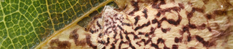
And two other upper and lower surface pairings from my photo archives; some of these look a lot more like leaf mines than did the circular things I asked Dave Wagner about 18 years ago:


These last four photos were all taken in New England in June. The “galls” in the last example are all already abandoned, even though there are four that appear only as faint squiggly impressions, without any of the purple or yellow discoloration, which as you can see in the previous examples is normally well developed by the time the larvae are done feeding. I have no explanation for this. Plenty still to be learned about this strange little fly!


