1. X
  2. NephroPOCUS
Log inSign up
NephroPOCUS
38.1K posts
Image
user avatar
NephroPOCUS
@NephroP
#POCUS #MedEd education 📖 | by Abhilash Koratala MD @KoraAbhi, “Echo-friendly” Nephrologist and Intensivist I Founding Member @POCUSIAPN | X≠ medical advice
nephropocus.com
Joined November 2018
307
Following
96.7K
Followers
RepliesRepliesArticlesArticlesMediaMedia

New to X?

Sign up now to get your own personalized timeline!

Create account

By signing up, you agree to the Terms of Service and Privacy Policy, including Cookie Use.

Terms·Privacy·Cookies·Accessibility·Ads Info·© 2026 X Corp.
Don't miss what's happening
People on X are the first to know.
Log inSign up
  • Pinned
    user avatar
    NephroPOCUS
    @NephroP
    Aug 22, 2022
    Time to update the #VExUS resources pinned thread 🧵📌 1/ Tweetorial on image acquisition. #POCUS #MedEd
    user avatar
    NephroPOCUS
    @NephroP
    Jun 8, 2022
    #POCUS #MedTwitter #Nephpearls Many #VExUS enthusiasts asked for a #tweetorial on image acquisition pearls. Did one b4 but time for an updated one 🧵 #1 Let's start with basics Color Doppler identifies the flow + tells the direction (blue is away & red towards the probe [BART])
    Image
    00:00
  • user avatar
    NephroPOCUS
    @NephroP
    Apr 26, 2023
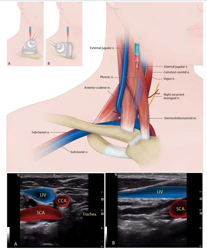
    Some excellent anatomic illustrations for friends who place central lines. #POCUS #MedTwitter #Nephpearls #FOAMed Courtesy: pubmed.ncbi.nlm.nih.gov/27521991/ 1. Internal jugular vein (A. Transducer placement in short axis, B. long axis)
    Image
    218K0218K
  • user avatar
    NephroPOCUS
    @NephroP
    Sep 4, 2022
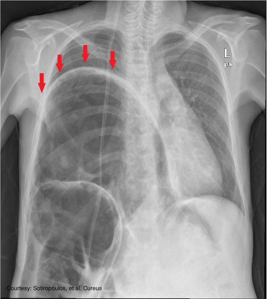
    A young woman recently diagnosed a periosteal femoral mass presented with dyspnea and pleuritic chest pain, and a chest x-ray was done. What is the likely diagnosis? Answer: 🔗nejm.org/doi/full/10.10… #MedEd #radiology #chestxray
    Image
  • user avatar
    NephroPOCUS
    @NephroP
    May 19, 2024
    #POCUS guided central venous cannulation illustrations #FOAMed #Nephpearls #FOAMcc
    Image
    00:00
    110K0110K
  • user avatar
    NephroPOCUS
    @NephroP
    Jun 25, 2024
    Comparison of Pros and Cons of #Dialysis Vascular Access Types. #MedEd #FOAMed Source: JAMA. 2024;331(15):1307-1317. PMID: 38497953.
    Image
    267K0267K
  • user avatar
    NephroPOCUS
    @NephroP
    May 17, 2023
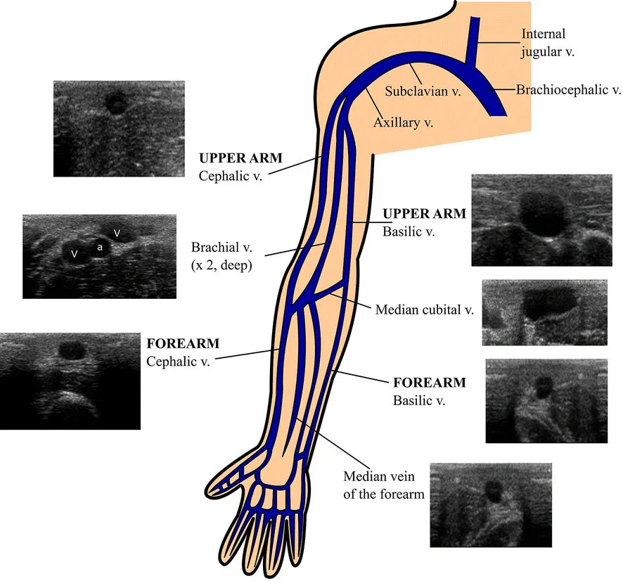
    Upper limb venous anatomy #POCUS #FOAMed #Nephpearls #MedEd theultrasoundjournal.springeropen.com/articles/10.11…
    Image
    208K0208K
  • user avatar
    NephroPOCUS
    @NephroP
    Oct 19, 2022
    What's happening here? #Xray #MedEd
    Image
  • user avatar
    NephroPOCUS
    @NephroP
    Dec 3, 2022
    #xray obtained from an elderly patient. What is this interesting finding? #MedEd #FOAMed Image courtesy: @DrGalenMD
    Image
  • user avatar
    NephroPOCUS
    @NephroP
    Oct 5, 2022
    What’s happening here? #xray #MedEd
    Image
  • user avatar
    NephroPOCUS
    @NephroP
    Jan 23, 2023
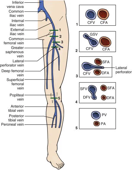
    Venous anatomy of the lower extremity. Click ‘ALT’ for figure description 🔗 atsjournals.org/doi/pdf/10.151… #POCUS #MedEd #Nephrology #FOAMcc
    The numbered points denote the minimum sites of compression for a limited examination. Note the paired common femoral artery, which divides proximal to the division of the common femoral vein (CFV), producing several cross-sectional segments of the CFV showing two adjacent arterial branches, the superficial femoral artery and the deep femoral artery. a = artery; v = vein.
    147K0147K
  • user avatar
    NephroPOCUS
    @NephroP
    May 16, 2023
    Lower extremity DVT #POCUS #anatomy radiologykey.com/lower-extremit… #IMPOCUS #FOAMed
    Image
    146K0146K
  • user avatar
    NephroPOCUS
    @NephroP
    Nov 18, 2022
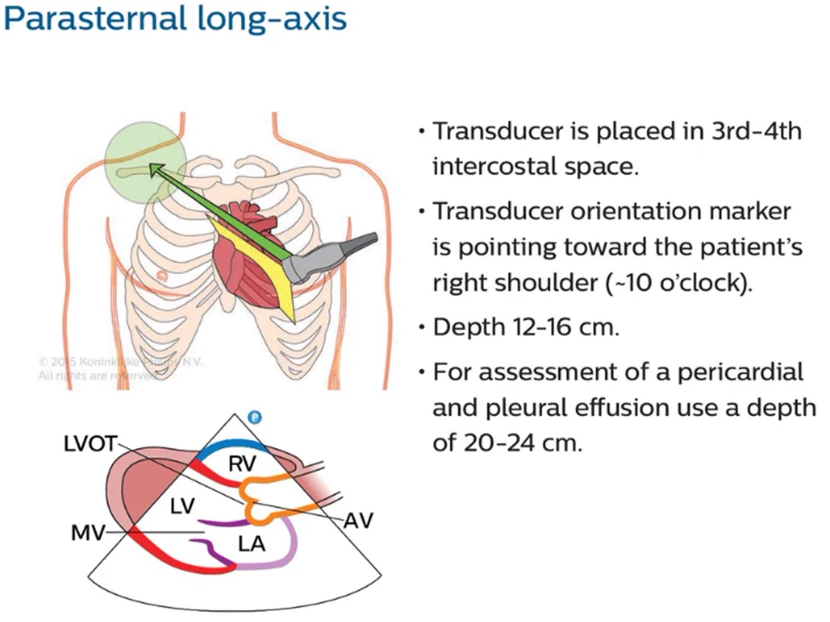
    Basic cardiac #POCUS views image acquisition flash cards. Courtesy of Philips Healthcare #MedEd #Nephpearls #echofirst 🧵 1/
    Image
  • user avatar
    NephroPOCUS
    @NephroP
    Jun 8, 2022
    #POCUS #MedTwitter #Nephpearls Many #VExUS enthusiasts asked for a #tweetorial on image acquisition pearls. Did one b4 but time for an updated one 🧵 #1 Let's start with basics Color Doppler identifies the flow + tells the direction (blue is away & red towards the probe [BART])
    Image
    00:00
  • user avatar
    NephroPOCUS
    @NephroP
    Aug 15, 2022
    Excellent #echofirst illustration depicting the relationship between cardiac cycle, Doppler parameters and M-mode at the mitral valve level. #MedTwitter #POCUS #Nephrology Courtesy🔗thoracickey.com/m-mode-imaging/
    Image
Advertisement
Advertisement