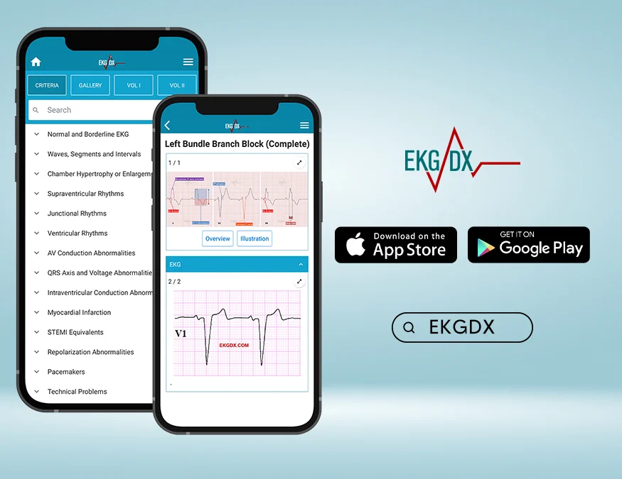
Let's talk about EKGDX App.
1/ Criteria
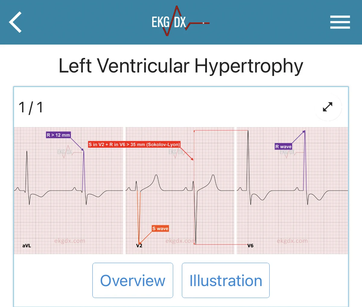
It contains the electrocardiographic criteria of each pattern deeply referenced, which allows you to read high-quality scientific content (+1500 scientific articles in the references) within the App.
🔗ekgdx.com#ecg#ekg
1/ Let’s talk about P waves #CardioTwitter.
The P wave is the first positive deflection on the EKG and represents atrial depolarization. The first half represents right atrial depolarization and the second half represents left atrial depolarization.
By @ekgdx
1/ Let’s talk about the ST segment #CardioTwitter.
ST segment normally represents the interval between ventricular depolarization and repolarization.
Normal ST
✅ Usually isolectric or may vary from 0.5 mm below to 1 mm above isolectric line in L leads.
#ekgdx#Medicine
1/ Let’s talk about the J wave #CardioTwitter.
The J wave is an extra positive deflection between the terminal portion of the QRS and the beginning of ST segment.
🧵👇by @ekgdxA
1/ Let’s talk about epsilon (ε) wave #Cardiotwitter
The epsilon wave can be defined as an electric signal of depolarization observed between the end of the QRS complex and the beginning of the T wave.
🧵👇 by @ekgdx
1/ Let’s talk about T waves #CardioTwitter
The T wave represents typically ventricular repolarization. It is the most labile wave on the EKG surface.
Normal T wave
✅ Morphology: Asymmetric.
✅ Amplitude: ≤6 mm in limb leads and ≤10 mm in precordial leads.
#MedTwitter
1/ Delta wave is slurring slow rise of initial portion of the QRS and is usually associated with short PR. It is most commonly associated with pre-excitation syndrome such as Wolff Parkinson White (WPW).
Delta waves can be:
✅ Positive
✅ Negative
by @ekgdx#CardioTwitter
1/ Let’s talk about PR Interval - Segment #CardioTwitter.
The PR interval represents the time between the onset of atrial depolarization and the onset of ventricular depolarization and reflects conduction through the AV node.
🧵by @ekgdx
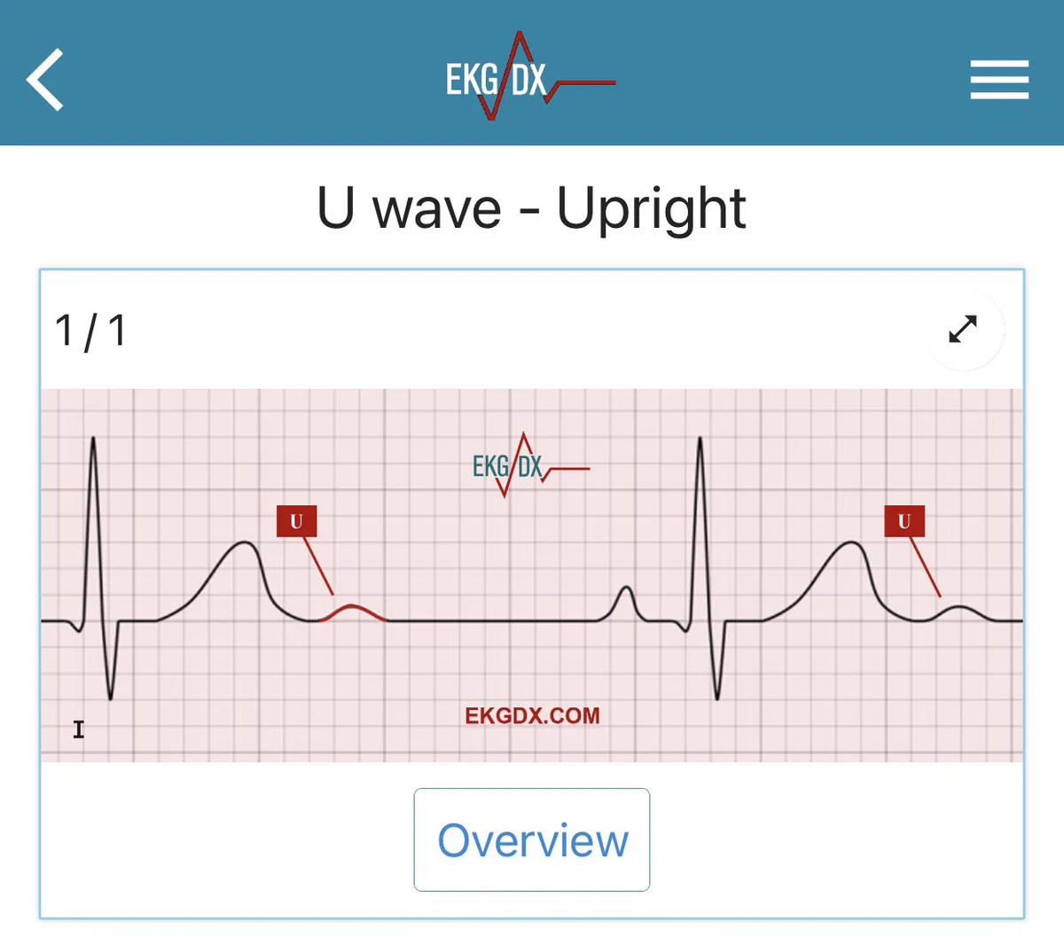
1/ Let’s talk about U waves #CardioTwitter.
The U wave is a deflection (usually small) immediately following the T wave.
The U wave can be:
✅ Positive: upright with amplitude <1.5 mm.
✅ Prominent: upright with amplitude >1.5 mm.
✅ Inverted or negative.
🧵👇 #ekgdx
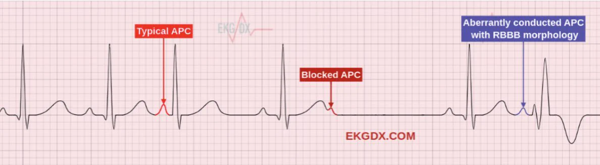
1/ Let’s talk about atrial premature complex (APC) #CardioTwitter.
APC occurs when an ectopic focus within the atria, generates an action potential before the next scheduled sinus beat and it is often characterized by an abnormal and premature P wave.
🧵👇by @ekgdx#ekgdx
Acute pulmonary embolism (PE) is one of the most serious form of venous thromboembolism. The clinical presentation of PE is variable and often nonspecific making the diagnosis challenging.
1/
#CardioTwitter