Log inSign up
Andreas Tolias Lab @ Stanford University
1,177 posts
Image
user avatar
Andreas Tolias Lab @ Stanford University
@AToliasLab
to understand intelligence and develop technologies by combining neuroscience and AI
Palo Alto, CA
toliaslab.org
Joined May 2017
819
Following
5,023
Followers
  • Pinned
    user avatar
    Andreas Tolias Lab @ Stanford University
    @AToliasLab
    Jun 17
    A hundred years ago, Edgar Adrian recorded from a single nerve fibre and showed the stimulus out in the world is carried in the rate of its firing — he started calling the impulses a "code." We've been trying to read that code ever since. For parts of the brain like V1 we've had
    user avatar
    Vedang Lad
    @vedanglad
    Jun 16
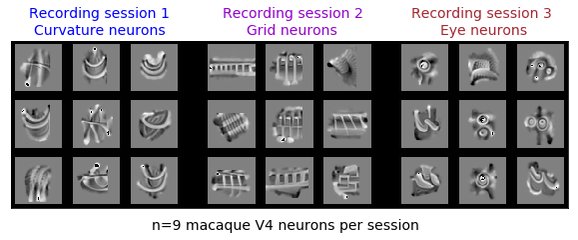
    How well can you describe the feature selectivity of a vision neuron … with words? Interpretability has long borrowed from neuroscience — and maybe it can give back too! 🧵
    Image
    00:00
    5.8K
  • user avatar
    Andreas Tolias Lab @ Stanford University
    @AToliasLab
    May 18, 2023
    Does the concept of cortical columns extend to higher-level primate cortex? Using #DeepLearning & physiology, we found that V4 neurons cluster in columns & form functional groups biorxiv.org/content/10.110… Led by @KonstantinWille @kelli_restivo w/ @sinzlab @kfrankelab @alxecker 🧵
    Image
    GIF
    176K
  • user avatar
    Andreas Tolias Lab @ Stanford University
    @AToliasLab
    Mar 27, 2020
    Functional connectomics to mechanistically dissect cortical algorithms. We align 2P calcium with EM data using CNMF framework to recover cell body and dendritic activity in EM volume. Thanks @IARPAnews for funding biorxiv.org/content/10.110…
    Image
    00:00
  • user avatar
    Andreas Tolias Lab @ Stanford University
    @AToliasLab
    Apr 11, 2025
    After 7 years, thrilled to finally share our #MICrONS functional connectomics results! We recorded activity from ~75K neurons in visual cortex in a single mouse, then mapped its wiring using electron microscopy. To systematically characterize neuron function, we built the first
    Image
    39K
  • user avatar
    Andreas Tolias Lab @ Stanford University
    @AToliasLab
    Oct 22, 2020
    Multimodal census-atlas of cell types of mouse and primate motor cortex including Patch-Seq data. Massive effort from biccn.org labs of @NIH BRAIN Initiative. Proud to be part of it with @CellTypist @FedericoScala7 @hippopedoid @sandberglab disq.us/t/3seoncm
    Image
  • user avatar
    Andreas Tolias Lab @ Stanford University
    @AToliasLab
    Mar 29, 2024
    Last day @bcm_neurosci after an incredible 15+ yrs. Immensely thankful to our brilliant students, postdocs, staff, collaborators, and colleagues. Next up: @Stanford! Thrilled for new adventures and collaborations in the Bay Area as we unlock the secrets of intelligence!
    31K
  • user avatar
    Andreas Tolias Lab @ Stanford University
    @AToliasLab
    Apr 24, 2020
    With @AllenInstitute and @SebastianSeung we release our MICrONS @IARPAnews phase 1 functional connectomics data microns-explorer.org. Movie shows 2P calcium and EM data aligned together using EASE with cell body and dendritic activity in EM volume. biorxiv.org/content/10.110….
    Image
    00:00
    Image
    user avatar
    allen institute
    @AllenInstitute
    Apr 24, 2020
    Announcing the MICrONs Explorer: A virtual observatory of the cortex 👉 microns-explorer.org This data portal includes #openscience #electronmicroscopy based reconstructions of cortical circuitry from mouse visual cortex.
  • user avatar
    Andreas Tolias Lab @ Stanford University
    @AToliasLab
    Nov 4, 2019
    #InceptionLoops combine large scale recordings with #DeepLearning to reveal a neuron's feature selectivity, such as most exciting inputs. Optimal stimuli for mouse V1 are not Gabors. nature.com/articles/s4159… in #NatureNeuroscience Lead by @eywalker @sinzlab together with @ecobost
    Image
  • user avatar
    Andreas Tolias Lab @ Stanford University
    @AToliasLab
    Nov 18, 2024
    Think government ROI is low? In the ‘60s, #NIH-funded researchers Hubel & Wiesel studied how cats’ neurons process visual info—seemed trivial, right? Yet their work led to convolutional neural networks like Hinton’s AlexNet, sparking the AI revolution that’s reshaping the global
    user avatar
    Vivek Ramaswamy
    @VivekGRamaswamy
    Nov 18, 2024
    The ROI for most government expenditures is very low.
    32K
  • user avatar
    Andreas Tolias Lab @ Stanford University
    @AToliasLab
    Oct 28, 2022
    1/4 Excited to share our latest work on feedback (FB). nature.com/articles/s4146…. There are abundant FB connections between cortical areas. To understand its function and its circuit level mechanisms we need to discover the FB cell type writing rules.
  • user avatar
    Andreas Tolias Lab @ Stanford University
    @AToliasLab
    May 18, 2024
    Check out our new #BCI shorturl.at/nKwnH. Subdural, flexible μECoG device features 65,536 channels, with bi-directional wireless communication and power enabling decoding the primate brain with high spatiotemporal resolution. Compare specs to other #BCIs
    Image
    28K
  • user avatar
    Andreas Tolias Lab @ Stanford University
    @AToliasLab
    Sep 16, 2019
    Canonical circuits are thought to makeup the #cortex. But what is canonical about them? Lead by @FedericoScala7 and Dmitry Kobak we show that not all cell types are present across areas and propose that specific circuit motifs are used as building blocks disq.us/t/3i8a85j
    Image
  • user avatar
    Andreas Tolias Lab @ Stanford University
    @AToliasLab
    Jul 30, 2021
    Proud to release our MICrONS multi-area data microns-explorer.org, the largest functional connectome to date, containing 200,000 cells, 75,000 neurons with functional imaging, and 523 million synapses. Excited to see the discoveries from this dataset.
    Image
  • user avatar
    Andreas Tolias Lab @ Stanford University
    @AToliasLab
    Nov 13, 2020
    1/2 As suggested by our huge Patch-seq effort lead by @FedericoScala7 and @hippopedoid published today in @nature the tree of cortical cell types seems more like a banana tree with a few large leaves, rather than an olive tree with many small ones disq.us/t/3sufdoi
    Image

New to X?

Sign up now to get your own personalized timeline!

Create account

By signing up, you agree to the Terms of Service and Privacy Policy, including Cookie Use.

Terms·Privacy·Cookies·Accessibility·Ads Info·© 2026 X Corp.
Don't miss what's happening
People on X are the first to know.
Log inSign up
Advertisement
Advertisement