Chris Appleton and I have a sad news to share with the entire #Echo@ASE360 community.Liv Hatle, the legend, pioneer, and mentor to ALL of us passed away on 6/23. When we saw her a mo. ago, she was talking about her new book and calcific MS. Let us celebrate her amazing career!
Thanks for sharing those fantastic IVC images and hepatic vein Doppler tracings. Hepatic vein Doppler taught by #LivHatle is critical for @AAE@ASE360 echo assessing different underlying pathologies for congestion as well as volume status. Should be a part of comprehensive Echo.
Another critical Echo feature of constrction is preserved (>= 8cm/s) or ⬆️ mitral Medial Annulus e' velocity which is ⬇️ in ALL MYOCARDIAL DISEASES. When CP is mixed with myopathy, e' is lower than that in pure CP. Constriction until proven otherwise when e' is⬆️ in HF patient!
#0/7 Thanks @purviparwani for robust discussion on diastology which means "Dilation".
I was asked to present my approach to Diastolic Function assessment at @ase360 " Just Relax: Diastolic Dysfunction" session. Let me share again and explain the 7 points that I emphasized.
Stuck in echo lab today but loving Dr. @JaeKOh2 talk on diastology! Thank you @ASE360 for live stream!
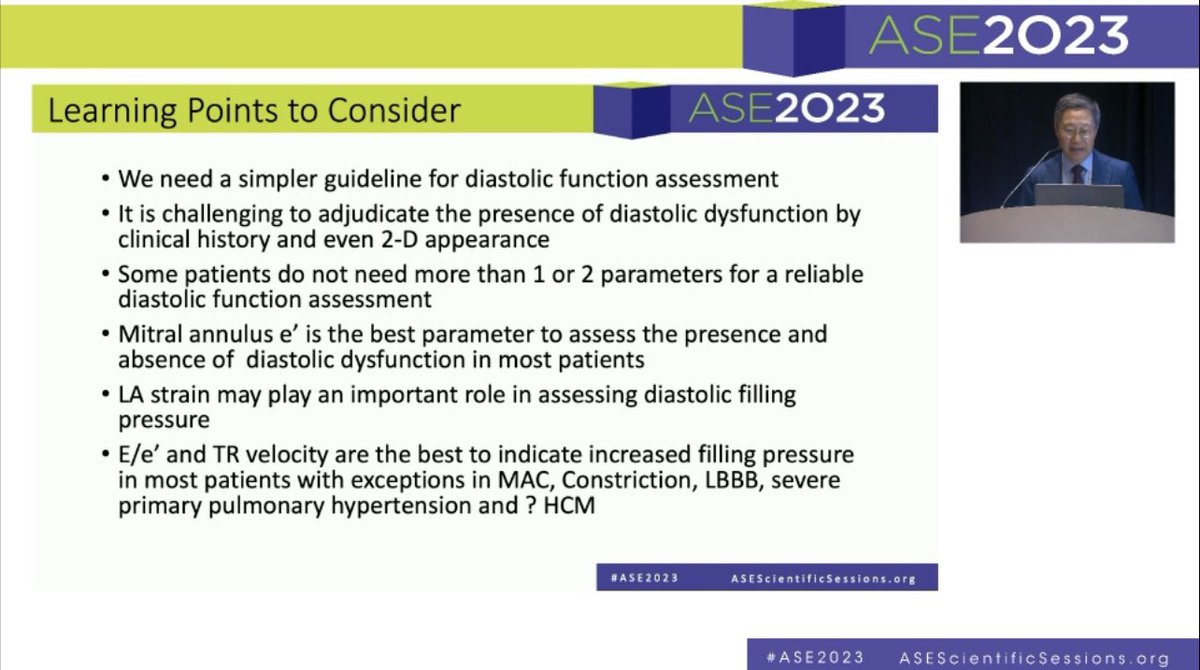
1. E prime is the most important parameter
2. Systolic dysfunction doesn’t always mean diastolic dysfunction
2. Not all the diastolic parameters are required
3. Variability
Thanks for tweeting this @HeartDocSharon who gave a great contrast talk @ASE360. Here is real time animation #LivHatle and we @MayoClinicCV put together 20 years ago. The best demonstration of constriction hemodynamics. Do not miss constriction which is a CURABLE Diastolic HF.
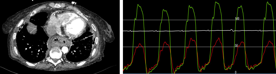
A 65 yo pt was referred to cardiac surgery for pericardiectomy with CT and cath (see below) consistent with constriction. Intraoperative @ase TEE was performed by an outstanding anesthesiologist. What do you think he said to the surgeon who is about to do sternotomy? @aae_echo
It is a myth that diastolic function is always abnormal in patients with systolic dysfunction. Young patients with reduced EF can have normal DF and filling pressure. Shown below was obtained from a 40 year old woman with LVEF of 25%. Filling pressure is normal. @jamil_tajik
For me that’s a problem, and there are several actually:
1. Diagnosing anyone with systolic dysfunction with diastolic dysfunction is pointless as by definition the two go together
2. In ICU loading conditions constantly change so what looks like grade 1 can actually be grade 2
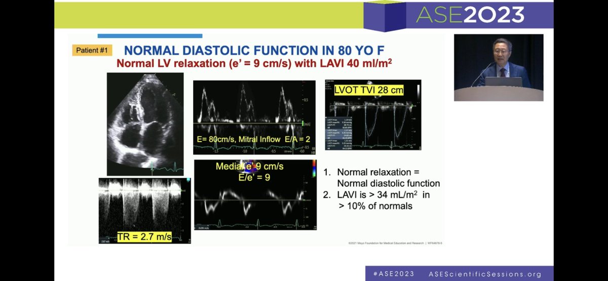
I was trying to emphasize that LV diastolic dysfunction cannot happen with normal myocardial relaxation. The best way to assess LV relaxation is mitral annulus e' velocity. So, medial or septal e' >=9 cm/sec indicates normal relaxation and diastolic function.@aae_echo@ASE360
1/5 My Dx for the e' velocity of 4 cm/s is reduced myocardial relaxation, which is sine qua non of diastolic dysfunction. It is present in all forms of myocardial disease and also with aging. Let me explain its progression & how to use the information for our pts @MayoClinicCV
This HV Doppler shows increased inspiratory diastolic flow reversal cw high RV DP due to a myocardial disease. The last cycle was with inspiration since forward flow velocity increased. Diastolic reversal happens with expiration in constriction. @jamil_tajik Not TR!
@EchoCases
1/3. @EleidMack asked me to review Echo which was interpreted as normal. Looking at closely, Echo shows a subtle septal motion change. M-mode of LV would have shown septal motion change more clearly.
Mitral inflow velocity showed >25% respiratory variation suggestive of CP.
1/2 Just received a box of chocolate from my pt who underwent pericardiectomy during COVID by @MayoClinicCVS after several yrs of HF elsewhere. Had numerous thoracenteses and liver evaluations including a biopsy. #Echo diagnosed CP using #Hatle and @MayoClinicCV dx criteria.
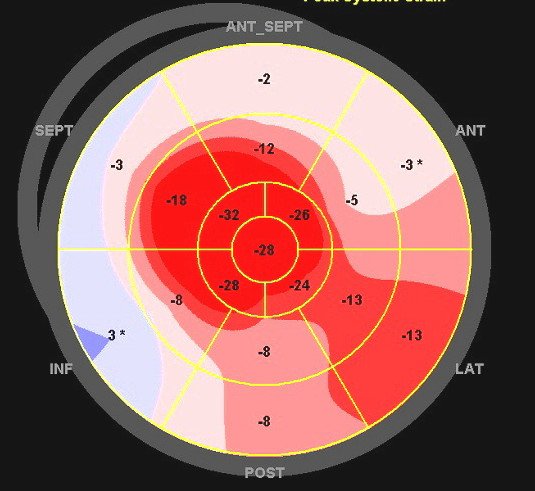
Doppler, Color M mode, and strain from a patient with HCM. A good example of short IVRT and "L" wave indicating increased filling pressure. E velocity flow propagation velocity does not work in a small LV as in HCM.@jamil_tajik @ASE360@aae_echo@KyleWKlarich@DavidWienerMD
From @MayoClinicCV#hemody session. PV premature diastolic opening is related to increased RVEDP, but not necessarily increased mean RV diastolic pressure. (See 2 examples below) Same in the LV. @jamil_tajik @ASE360@aae_echo@EchoCases