korbinhaycockmd
8,782 posts
Image
user avatar
korbinhaycockmd
@khaycock2
POCUS, NBE CCE diplomat, Echo, Resuscitation, VExUS, Emergency Medicine, 🏴
Joined February 2019
306
Following
5,107
Followers
  • user avatar
    korbinhaycockmd
    @khaycock2
    May 29, 2022
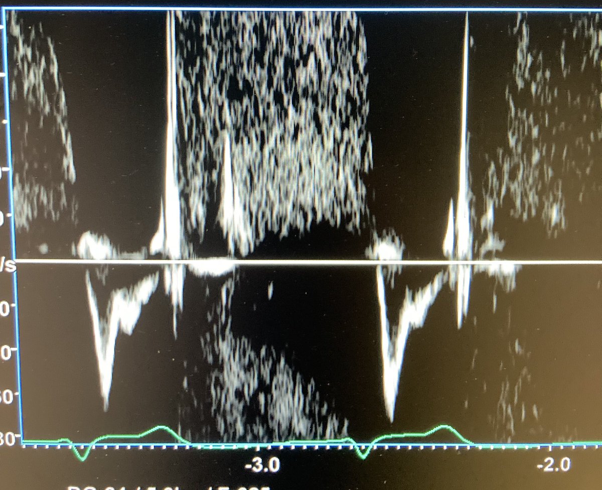
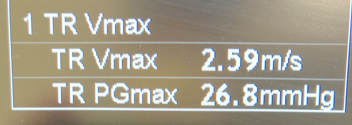
    Trauma patient due to MVA. HR 50, sinus rhythm, BP 51/32 & confused. Due to hypotension & trauma, trauma team is activated prior to arrival. eFAST normal except for the image on the left. Epinephrine gtt started and HR & BP normalized. A complex situation is easy c #POCUS
    Image
    00:00
  • user avatar
    korbinhaycockmd
    @khaycock2
    Feb 6, 2022
    1/ A patient with massive pulmonary embolism given tPA. After tPA, still severe RV dysfunction. RVOT Doppler:
    Image
  • user avatar
    korbinhaycockmd
    @khaycock2
    Mar 1, 2022
    1/ 44 yo patient came to ED with SBP 70 mmHg, HR 150-160 atrial fib. Was s/p ablation for atrial fib 12 days ago and had sudden presyncope and palpitations. POCUS: plethoric IVC and reverse S on HV,diffuse B-lines, TRpv 35 mmHg (so sPAP about 55-60), & the following other images:
  • user avatar
    korbinhaycockmd
    @khaycock2
    Mar 20, 2021
    Can anyone, in any situation, ever rationalize giving 24 L of fluid in 24 hours? This actually happens.
  • user avatar
    korbinhaycockmd
    @khaycock2
    Oct 18, 2021
    1/ A case of increasing the cardiac output by manipulating the arterial elastance/afterload rather than using an inotrope:
  • user avatar
    korbinhaycockmd
    @khaycock2
    Jan 26, 2022
    65 y/o patient. Brought in as trauma because of syncope with hypotension despite IVF by medics. eFAST is done right after arrival. While standing over the resident’s shoulder, RV is noted to be large with septal shift. Doppler of the TV and RVOT VTI is immediately done:
    Image
    Image
    Image
  • user avatar
    korbinhaycockmd
    @khaycock2
    Jan 2, 2024
    Cardiogenic shock and how serious it is is under appreciated because often the BP is normal. A symptomatic patient with a CO only supported by tachycardia is a medical emergency. A MAP generated by CVP and SVR with a low SV and tachycardia to keep a CO just hanging in=danger.
    user avatar
    IMCrit
    @IM_Crit_
    Jan 1, 2024
    ICU Cheat Sheet for Management of Acute-on-Chronic Heart Failure Complicated by Cardiogenic Shock:
    Image
    14K
  • user avatar
    korbinhaycockmd
    @khaycock2
    Aug 20, 2021
    1/ TCAV as a ventilator strategy in the Emergency Department? Here’s a case: A patient with COVID intubated for hypoxemic respiratory failure. O2 sats were mid 70% on 100% CPAP prior to intubation with persistent & significant increased WOB. Here’s the CXR after intubation:
    Image
  • user avatar
    korbinhaycockmd
    @khaycock2
    Aug 22, 2021
    1/ Massive PE patient with high clot burden and saddle embolism. RVOT VTI before milrinone/epoprostenol medneb:
    Image
  • user avatar
    korbinhaycockmd
    @khaycock2
    Aug 30, 2022
    1/ A patient presents with encephalopathy and SOB. Edematous, cold and clammy. Initial bedside echo with SV 48 cc and CI 1.6 L/min/m2. Pre-ejection time 97 msec and total ejection time 290 msec.
    Image
    Image

New to X?

Sign up now to get your own personalized timeline!

Create account

By signing up, you agree to the Terms of Service and Privacy Policy, including Cookie Use.

Terms of Service|Privacy Policy|Cookie Policy|Accessibility|Ads info|© 2026 X Corp.
Don't miss what's happening
People on X are the first to know.
Log inSign up
Advertisement
Advertisement