Log inSign up
Divya Gunda
579 posts
Image
user avatar
Divya Gunda
@learnneurorad
Neuroradiologist @CooperRadRes, here to share #Neurorad cases for #radres #RadEd. Tweets my own, for #MedEd, not medical advice. Alum: @PennRadiology @OUHealth.
instagram.com/learnneurorad
Joined September 2015
432
Following
5,888
Followers
  • user avatar
    Divya Gunda
    @learnneurorad
    Jul 16, 2022
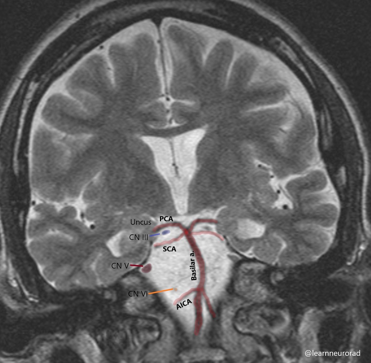
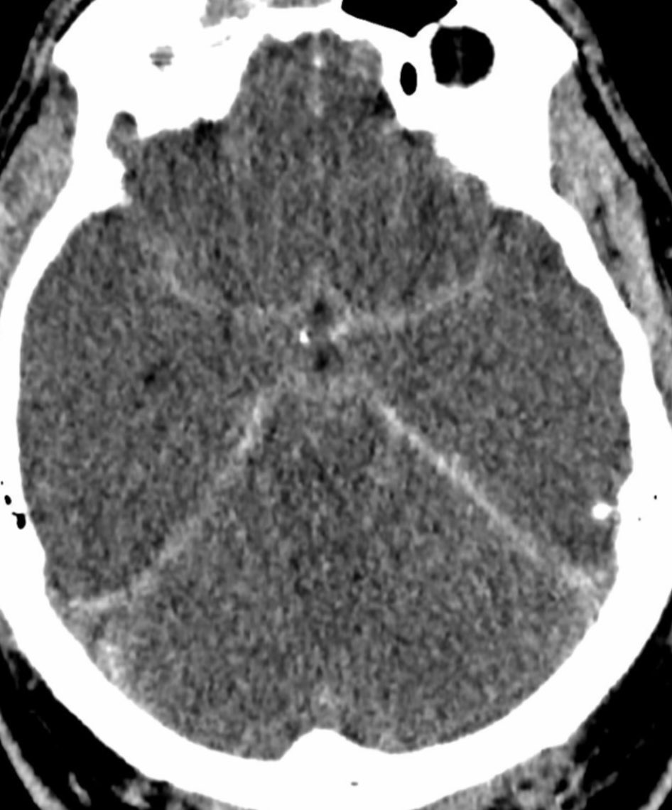
    Cranial nerve anatomy to help understand why uncal herniation and Pcomm aneurysms result in a fixed and dilated pupil. CN III and IV run b/w the PCA & SCA. CN V and VI run in the prepontine cistern, lateral to the basilar artery. #Neurorad #radres #Neurotwitter #MedEd #MedTwitter
    Image
  • user avatar
    Divya Gunda
    @learnneurorad
    Dec 22, 2021
    When to call normal vs. volume loss vs. normal pressure hydrocephalus vs. chronic compensated hydrocephalus. #radres #NeuroRad #Neurotwitter #Neurosurgery #Neurology #MedEd #neuroscience #FoamRad #FOAMed
    Image
  • user avatar
    Divya Gunda
    @learnneurorad
    Jan 20, 2022
    Basal cistern anatomy on an axial CT of the head. Perimesencephalic cistern is composed of the paired crural and ambient cisterns and the unpaired interpeduncular and quadrigeminal cisterns. #NeuroRad #radres #NeuroTwitter #Neurosurgery #MedEd #neuroanatomy #FOAMrad #MedTwitter
    Image
  • user avatar
    Divya Gunda
    @learnneurorad
    Nov 15, 2022
    Do not mistake pars intermedia cysts for pituitary adenomas. They are a common incidental finding, with simple or proteinaceous cyst content, located in between the pars distalis and pars nervosa, without mass effect or suprasellar extension. #radres #Neurorad #Neurosurgery #nsgy
    Image
  • user avatar
    Divya Gunda
    @learnneurorad
    Feb 10, 2022
    The radiologist may often be the first to diagnose idiopathic intracranial hypertension. Diagnosis can be challenging as these patients can have normal opening pressures. Look for these findings on MRI, which are not always present together. #Neurorad #radres #NeuroTwitter
    Image
  • user avatar
    Divya Gunda
    @learnneurorad
    Sep 7, 2024
    More anatomy of the jugular foramen from #ASHNR24. The jugular foramen is divided into the pars nervosa and the pars vascularis by the jugular spine. #radres #Neurorad
    Image
    40K
  • user avatar
    Divya Gunda
    @learnneurorad
    Jan 27, 2022
    While there are many classification systems for ICA anatomy, the NYU classification seems to be more practical on cross sectional imaging and removes some confusion regarding the lacerum and clinoid segments. #radres #Neurorad #NeuroTwitter #Neurosurgery #MedEd #FOAMrad
    Image
  • user avatar
    Divya Gunda
    @learnneurorad
    Apr 21, 2022
    While we don’t routinely assess for ligamentous injury on CT, it’s important to look at the soft tissue windows on CT to look for gross disruption of these normal craniocervical junction ligaments. #Neurorad #radres #Neurotwitter #Neurosurgery #Spine #Orthotwitter #MedEd #nsgy
    Image
    Image
    Image
  • user avatar
    Divya Gunda
    @learnneurorad
    Apr 23, 2023
    Difference b/w SAH (left): preserved gray-white differentiation. No ventricular/sulcal effacement Pseudo-SAH (R): typically seen in diffuse cerebral edema/hypoxic ischemic injury caused by hyperdensity of arteries/veins relative to the low attenuation of brain #Neurorad #radres
    Image
    Image
    39K
  • user avatar
    Divya Gunda
    @learnneurorad
    Sep 28, 2022
    Several sites of intracranial calcifications can be mistaken for pathology. Note sites of physiologic calcifications in adults. Not as dense choroid plexus Ca2+ in the foramen of Monro can be mistaken for a colloid cyst. #radres #Neurorad #neurotwitter #Neurosurgery #MedTwitter
    Image
  • user avatar
    Divya Gunda
    @learnneurorad
    Jan 14, 2023
    Use the snail eyes sign to locate the facial nerve on a coronal CT of the temporal bone. Start with finding the snail shell/cochlea. The eyes reflect two segments of the facial nerve and the nose is the tendon of the tensor tympani muscle. #Neurorad #anatomy #radres #MedTwitter
    Image
    32K
  • user avatar
    Divya Gunda
    @learnneurorad
    Sep 21, 2022
    Dilated perivascular spaces can be confused for lacunar infarcts in the basal ganglia. More of an issue on CT than on MRI. Key points to help differentiate ⤵️ #Neurorad #radres #FOAMrad #neurotwitter
    Image
  • user avatar
    Divya Gunda
    @learnneurorad
    Dec 29, 2021
    Locating these skull base foramina starts with the anterior clinoid process. Optic canal is separated from superior orbital fissure by optic strut. Foramen rotundum (FR) is inferior to SOF and vidian canal is inferomedial to FR in the floor of the sphenoid sinus #NeuroRad #radres
    Image
  • user avatar
    Divya Gunda
    @learnneurorad
    Sep 12, 2024
    Normal anatomy of the lumbar spine #radres #Neurorad
    Image
    30K

New to X?

Sign up now to get your own personalized timeline!

Create account

By signing up, you agree to the Terms of Service and Privacy Policy, including Cookie Use.

Terms of Service|Privacy Policy|Cookie Policy|Accessibility|Ads info|© 2026 X Corp.
Don't miss what's happening
People on X are the first to know.
Log inSign up
Advertisement
Advertisement