#EPeeps How to distinguish in #ECG between fascicular PVC and PVC from papillary muscle❓..
Both present with a RBBB morphology❗️But..
Pap.muscle➡️longer QRS duration;R>r' QRS morphology in V1
Fascicular➡️shorter QRS; r<R' in V1
Why? See the illustration below!
#cardiotwitter
#EPeeps here is our #EP_Kiel@evgeny_lyan illustration for Coumel Law! Based on incredible publication doi.org/10.1161/CIRCUL…
If a wide complex tachy➡️narrow complex tachy with ⬆️HR- this is orthodromic AVRT with AP on the same side as the blocked bundle branch! #CardioTwitter
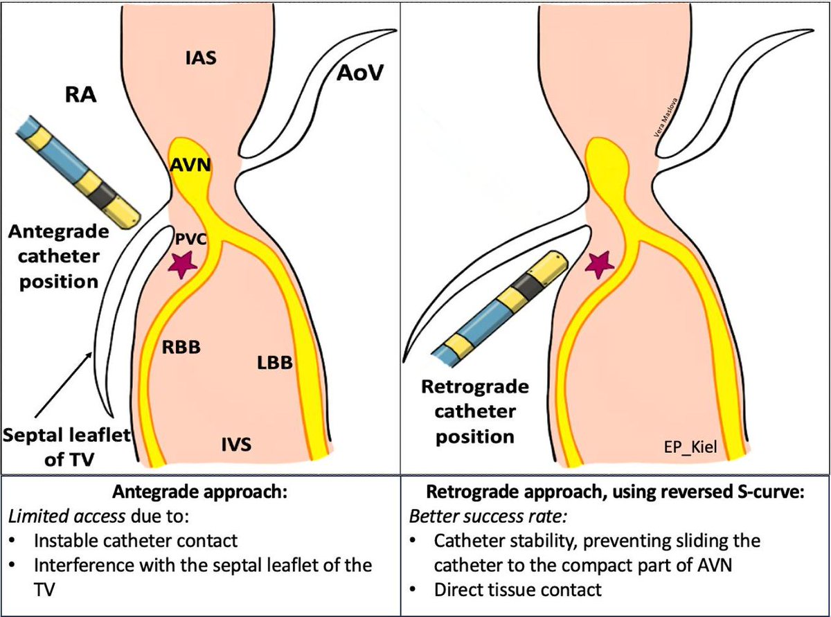
#EPeeps Why do we use reversed S- curve for parahisian PVC ablation? @evgeny_lyan
✅catheter stability under the TV leaflet
✅direct tissue contact
#EP_Kiel#Cardiotwitter
AP from the last case- LEFT ANTEROLATERAL.
All we need for this diagnosis is in the 1Step of Arruda Algorithm:
Polarity of delta wave:
-Lead I isoelectric/even negativ(+-)➡️left free wall
-Lead aVF positiv(+)➡️anterolateral
Next post- mapping!
#Epeeps#CardioTwitter#ECG
#EPeeps DONT be fooled by P wave morphology! Check out the case from our lab with huge slow conducting area between AT origin and exit site #cardiotwitterx.com/ThomasDemming/…