Beautifully colorful #microscopy image of chameleon embryo from a #ZEISS system @MBLScience embryology course by Teresa Zgoda.
ZEISS Microscopy
10.6K posts
We are #Microscopy. All about microscopes from #ZEISS.
LinkedIn: zeiss.ly/ln Facebook: zeiss.ly/fb | Imprint & Data Privacy: zeiss.com
- Today, we announce the winners of the ZEISS Microscopy Image Contest 2021! 🏆🔬 ZEISS users from around 50 countries submitted more than 1,300 fascinating entries. A big thank you to all the participants! Discover the four winning images in this thread 👇 #ZEISSImageContest
- Boo! Happy #Halloween! 👻🎃😱 This image of a scared embryonic zebrafish face, tiled 20 times, was acquired with ZEISS LSM 800 confocal microscope. #ZEISS #Microscopy #Zebrafish #ZebfrafishZunday #microscreepy #spooktober
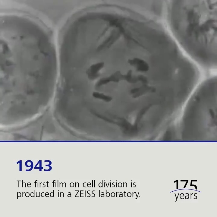
- The first film on cell division is produced in a ZEISS laboratory in 1943 – with the aid of a phase-contrast microscope. Just seven years earlier, the first prototype of a phase-contrast microscope was built at ZEISS in Jena. #ZEISS175 #throwback #celldivision
00:00 - Welcome to the fascinating world of gems, crystals, rocks and stones 💎⛏️: These four micrographs were acquired by ZEISS customers throughout the year 2021! 🙏🔬 (1/6) #zeiss #zeissmicroscopy #rawmaterials #geoscience #mineralogy
- Check out this cool #microscopy image Nipam Patel captured @MBLScience embryology course using a #ZEISS system!
- Another amazing image by Tessa Montague @MBLScience embryology course using a #ZEISS #microscope. This time a picture of a spider embryo!
- With this nanosized companion, known for his bravery, we celebrate yet another #StarWarsDay. May the 4th be with you!💫 #zeiss #microscopy #materialscience #nanosciencewithzeiss #nanofabrication #nanotech #MayThe4thBeWithYou
- Marine #plankton are the source of nearly all life in the sea. @PlanktonPundit uses his ZEISS zoom microscope to capture stunning images and movies of plankton. We are happy to support his fascinating work to make this hidden world visible: blogs.zeiss.com/microscopy/new… #WorldOceanDay
00:00 - It is official 🎉 The ribbon was cut a few moments ago. Now @AMI_microscopy Imaging Centre in collaboration with @afribioimaging and @ImagingAfrica will be able to empower the #microscopy community in Africa better than ever before. Here is a video #sneakpeek of the facility.
00:00 - What if you could quantify the mineralogy and its relationships, without sample preparation, to reveal all the internal structures of your sample? Discover our breakthrough innovation in #mineralogy zeiss.ly/t_mineralogic3D #geology #petrology #mining #geometallurgy #geoscience
00:00 - With these scary skulls – a.k.a. bamboo imaged with a ZEISS Sigma electron microscope – we wish you Happy Halloween and a spooky night! 💀👻 🔬 Image courtesy: Chenglin Xiao, Guangzhou, China
- Long-term volumetric imaging meets subcellular resolution. Join us on December 1: zeiss.ly/t-lattice-ligh… Image generated using AICS-0013 (laminB1-mEGFP) from the Allen Institute for Cell Science, Seattle, USA. @AllenInstitute #latticelightsheet #llsm #lls #microscopy
00:00Let's talk Lattice Lightsheet | Join us on December 1From zeiss.com


